VaRMI-CL
- Valvular Radiologic Multi-Imaging Core Laboratory (VaRMI-CL)
- Services and Information
- Service Request
Valvular Radiologic Multi-Imaging Core Laboratory (VaRMI-CL)
![]() The Valvular Radiologic Multi-Imaging Core Laboratory (VaRMI-CL) provides thorough analysis of heart valve images from different technologies such as axial tomography (CT scan), magnetic resonance imaging (MRI) and positron emission tomography (PET).
The Valvular Radiologic Multi-Imaging Core Laboratory (VaRMI-CL) provides thorough analysis of heart valve images from different technologies such as axial tomography (CT scan), magnetic resonance imaging (MRI) and positron emission tomography (PET).
Services and Information
Services offered by the VaRMI-CL:
- Transfer of images by a SFTP site
- Management and archival of data on a secure server with regular backups
- Numerous analysis software applications
- High-performance computing capabilities
- In-depth analyses, with quality assurance and standardization of image acquisition and interpretation
The VaRMI-CL provides image analysis services for research. For more information on undertaking the examinations themselves, visit the Advanced Imaging Platform (AIP) page or contact us for assistance in planning your project.
Application of Axial Tomography (CT Scan)
Axial tomography is useful in the diagnosis of valvular heart diseases. It allows for the quantification of cardiac calcifications, such as that of the aortic valve, mitral ring, coronary arteries or ascending aorta.
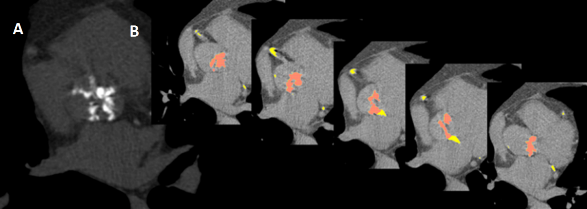
Calcification of the aortic valve seen by axial tomography
Measurement of calcium score is a marker for aortic stenosis severity. This measure also makes it possible to predict the occurrence of hemodynamic deterioration in aortic bioprostheses.
The injection of contrast agent during axial tomography permits viewing of non-calcified tissue. Assessing the above-mentioned severity or the degree of thrombosis/fibrosis in bioprotheses may then be refined.
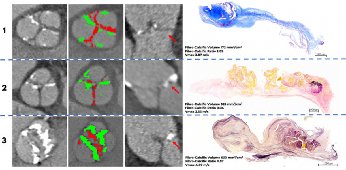
Contrast-enhanced CT and histology of aortic valve leaflets
Application of Positron Emission Tomography (PET) Imaging
Positron emission tomography is still being used as a research tool for valvular diseases. It enables visualization of active cellular-level mechanisms such as microcalcification, being one of the lesions responsible for the aortic stenosis to onset.
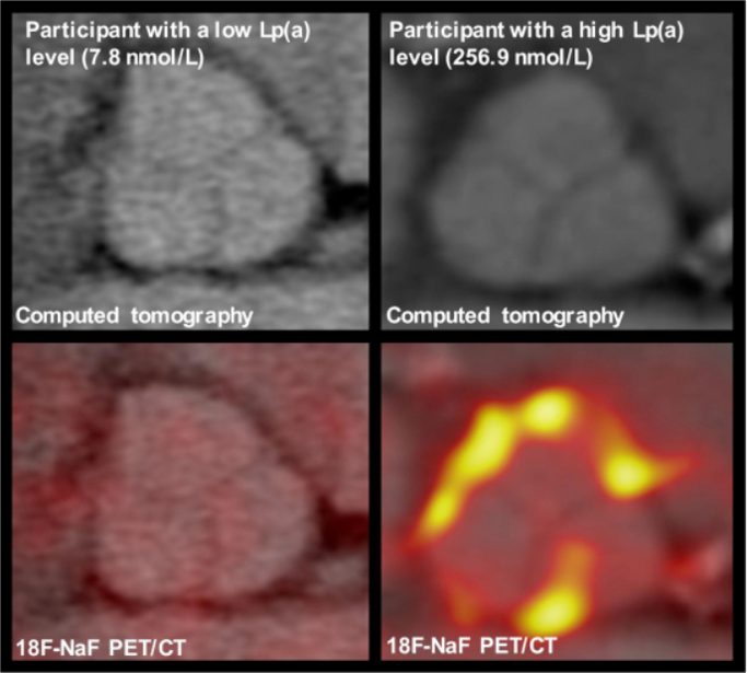
Microcalcification of the aortic valve seen by positron emission tomography
Completed or ongoing projects
- RESILIENCE Trial: Evaluation of the Durability of Aortic Bioprostheses/Valves with RESILIA Tissue in Subjects Under 65 - RESILIENCE (NCT03680040)
- Angiotensin Receptor Blockers in Aortic Stenosis - ARBAS (NCT04913870)
- Recherche sur les effets respiratoires du vapotage
- Diet Quality and Coronary Artery Calcification in Adults with Heterozygous Familial Hypercholesterolemia
- Using Premature Ventricular Contraction hemodynamics from the cardiac catheterization lab to discrIMINATE pseudo from true aortic stenosis – PVC-iminate Trial
- Contrast-Enhanced Computed Tomography Assessment of Aortic Stenosis. (Cartlidge et al. Heart. 2021)
- Association of Bioprosthetic Aortic Valve Leaflet Calcification on Hemodynamic and Clinical Outcomes (Zhang et al. J Am Coll Cardiol. 2020)
- Correlates of Coronary Artery Calcification Prevalence and Severity in Patients with Heterozygous Familial Hypercholesterolemia (Drouin-Chartier et al. CJC Open. 2020)
- Lipoprotein(a), oxidized phospholipids, and aortic valve microcalcification assessed by 18F-NaF PET/CT (Després et al. CJC Open. 2019)
- Genetic Variation in LPA, Calcific Aortic Valve Stenosis in Patients Undergoing Cardiac Surgery, and Familial Risk of Aortic Valve Microcalcification (Perrot et al. JAMA Cardiol. 2019)
- Sex-Related Discordance Between Aortic Valve Calcification and Hemodynamic Severity of the aortic stenosis: Is the Valvular Fibrosis the Explanation? (Simard et al. Circ Res. 2017)
Service Request
In order to get the cost assessment for your project, make a demand for services, or for any additional information, please contact:
Dre Marie-Annick ClavelDirector of VaRMI-CL
Marie-Annick.Clavel@criucpq.ulaval.ca